Dots in vision: the "glow" in the retina will help identify eye diseases at an early stage
- Статьи
- Science and technology
- Dots in vision: the "glow" in the retina will help identify eye diseases at an early stage


Russian researchers have found that changes in the "glow" of a special substance in the retina can detect its damage at the earliest stages — long before the appearance of serious visual impairment. Experts have studied a pigment that accumulates in epithelial cells with age and is able to luminesce under the influence of light: according to the characteristics of this glow, one can judge the condition of the eye. Experts interviewed by Izvestia noted that the technique will make it possible to diagnose pathologies at a stage when the development of the disease has not yet become irreversible, however, if negative changes are detected, patients will need to adjust their lifestyle.
Diagnosis of retinal diseases at the cellular level
Retinal diseases, including age-related degeneration, which is experienced by a large number of elderly people, are often detected in the late stages, when vision can no longer be restored.One of the reasons is the limited capabilities of existing diagnostic methods: they detect structural changes, but not early functional failures in cells.
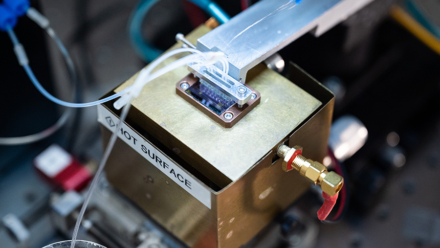
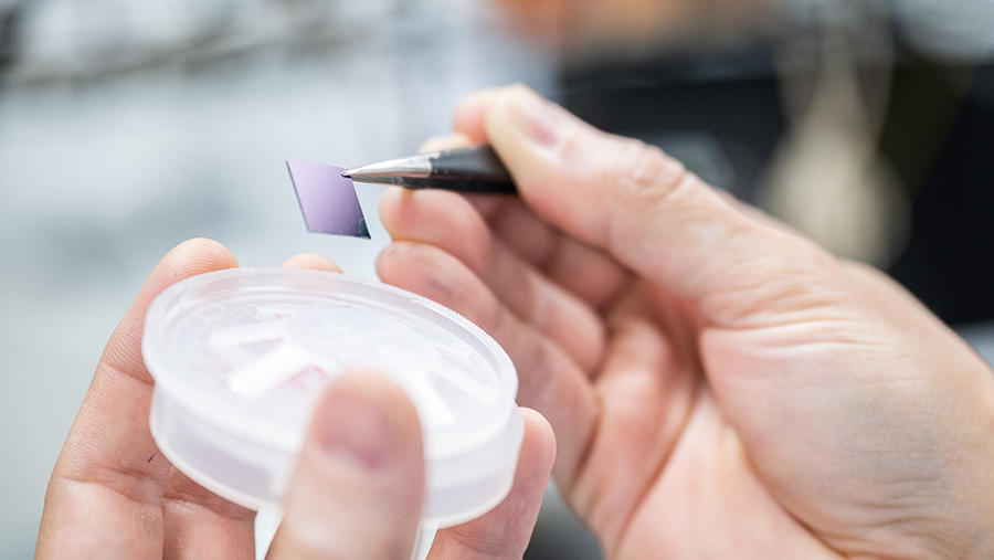
Scientists from NUST MISIS, Lomonosov Moscow State University, Moscow State University, MIPT, and the Institute of Bioorganic Chemistry named after Academicians M.M. Shemyakin and Yu.A. Ovchinnikov studied lipofuscin— a pigment that accumulates with age in cells of the retinal pigment epithelium, that is, the layer of epithelial cells that is located outside the nervous part of the retina. This substance is able to luminesce under the influence of light, and its characteristics can be used to judge the condition of the eye.
"The developed method for diagnosing retinal pathologies by the "glow" of cells will become an important tool for detecting diseases and evaluating the effectiveness of therapy," Alevtina Chernikova, rector of NUST MISIS, told Izvestia.
An important feature of lipofuscin is its phototoxicity: when exposed to visible light, it can generate reactive oxygen species and toxic oxidation products that cause pronounced oxidative stress. The study of these processes is important for understanding the mechanisms of retinal damage and diagnosing age-related degenerative changes. The ability of lipofuscin to autofluorescence plays an important role here. Measuring the parameters of its "glow" is an important tool for early diagnosis of eye diseases.
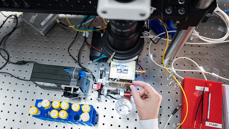
According to scientists, until now there was a lack of data on exactly how the composition of lipofuscin changes during light damage and how this affects the signal of its "glow". For the first time, Russian researchers have used the fluorescence lifetime imaging method to trace at the cellular level how a substance changes during oxidation inside cells of the pigment epithelium.
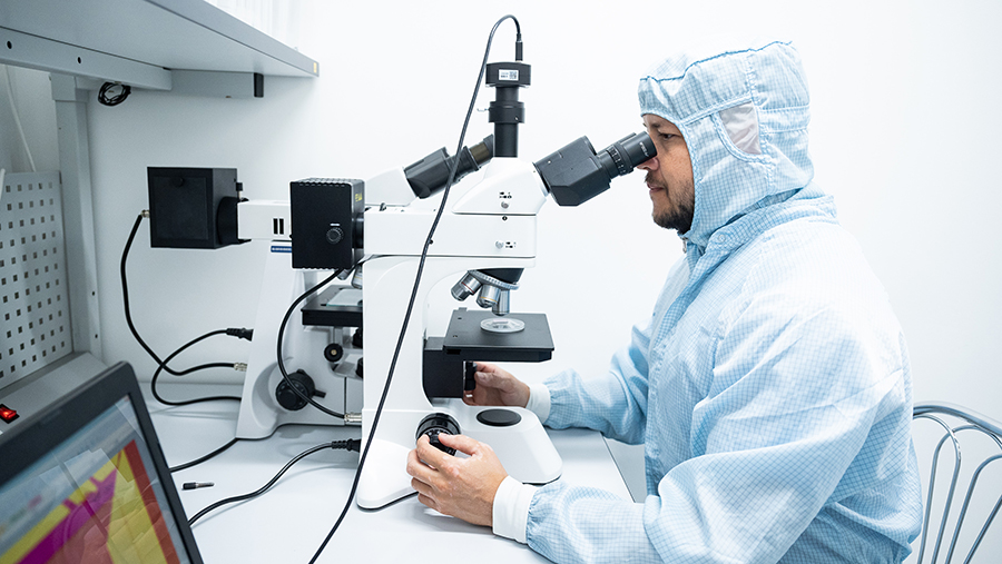
— It is important that we were able to detect these changes without introducing additional tags or interfering with the cell. Such measurements were made possible by fluorescence lifetime visualization. This is a modern microscopy method based on measuring the lifetime of the excited state of molecules, which allows us to obtain additional diagnostic information about the state of tissues," said Alexey Semenov, a researcher at the Laboratory of Photonic gas sensors at NUST MISIS.
The work was carried out under the program for attracting talented young scientists under the age of 39 within the framework of the Priority 2030 program.
Prospects for a new diagnostic approach
The scientists also analyzed the role of antioxidants in suppressing the phototoxic effects of lipofuscin. As part of the experiment, they studied the carotenoid protein AstaP, isolated from the microalgae Coelastrella astaxanthina and capable of delivering zeaxanthin, a natural substance that protects cells from oxidative stress. The study showed that such a complex slows down the destruction of lipofuscin: the formation of oxidized products decreases and complete damage to the pigment does not occur. The details of the work are published in the scientific journal Journal of Physical Chemistry B.
"In order to increase the sensitivity and speed of measurements, at the next stage we plan to use the quantum sensors we have developed — superconducting single—photon detectors," Grigory Goltsman, a leading researcher at the NTI MISIS Laboratory of Quantum Communications, shared his plans.
The main advantage of the proposed approach is the transition from structural diagnostics, when the doctor sees already formed druses, that is, deposits of eye waste products, or atrophy, to functional ones. This is the stage at which the process is still reversible, Marina Chumakova, a leading market expert at NTI Helsnet, told Izvestia.
— The method used by the authors is more objective than the standard autofluorescence of the fundus, which shows the intensity of the glow (dim/bright). It subjectively depends on the power of the device and the transparency of the media. As for the use of luminous proteins in general, the use of endogenous fluorophores makes it possible to obtain diagnostic information without introducing genetically modified structures or toxic dyes," the specialist said.

The work shows the possibility of detecting in the laboratory the accumulation of toxic forms of the pigment lipofuscin, which occurs in the eyes under prolonged illumination, explained Nikolai Ilyinsky, senior researcher at the Laboratory of Aging and Age-related Neurodegenerative Diseases at the Center for Research on Molecular Mechanisms of Aging and Age-Related Diseases at MIPT. The proposed procedure will make it possible to diagnose problems at an early stage without using additional contrast agents, which will accelerate treatment, including by correcting habits.
— After all, what suffers in the eye in the first place with the accumulation of photo damage? Supporting cells designed to recycle photoreceptor debris. And normalization of metabolism, that is, metabolism in the body as a whole, will help to strengthen the neutralization of toxic accumulations," the expert noted.
According to him, the accumulation of such damage manifests itself with age, when the efficiency of processing cellular "garbage" decreases, including due to bad habits. At the same time, new diagnostic methods will allow patients to pay attention to possible risks earlier and be more aware of their health.
Переведено сервисом «Яндекс Переводчик»
